
Early stage researchers in BioMEP doctoral programme
Golestan Karami: Study of brain tumor characteristics using advanced MR diffusion techniques and modeling

Primary supervisor: Professor Cosimo Del Gratta
Gabriele D’Annuzio University, Chieti-Pescara, Italy
Email: golestan.karami (at) gmail.com
Ongoing research in brain tumor imaging attempts to develop, validate, and clinically implement advanced neuroimaging techniques that result in radiological specification and grading which helps to plan and navigate surgery intra-operatively, to assess treatment, and prognosis.
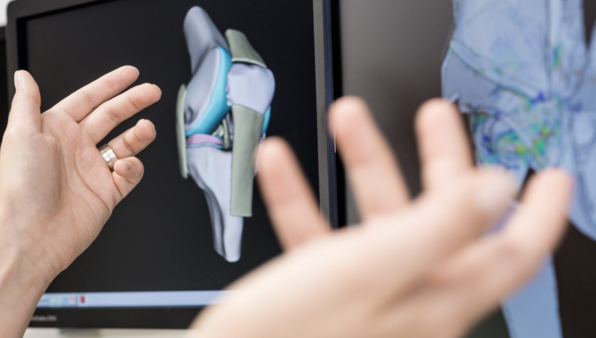
Diffusion Tensor Imaging (DTI) is a technique which is able to investigate structural and anatomical features of white matter in human subjects noninvasively. In fact DTI enables the measurement of the restricted diffusion of water in tissue in order to produce neural tract images, then having a great radiological value in disease where fibers are distorted (brain tumor).
Anisotropic diffusion is an effect of the microstructural properties of the voxel, and its change is a common feature of neuronal abnormalities and structural disorder. Recent studies have shown that Fractional Anisotropy (FA) value can help in understanding of the effect of brain tumors on white-matter fibers and the delineation of tumor margins beyond what is currently demonstrated with conventional imaging.
The main objective of the project is to investigate tissue microstructures and brain anatomy in brain tumor patients by using Diffusion technique and its potential benefits for identifying different tumor components, distinguish between tumor invasion from normal brain tissue or edema.
Another aspect of this research is to investigate quantification through the image processing with the possibility of developing new methods and assessment of the contributions of this type of imaging for surgery, early diagnosis or even evaluate patients after treatment and tumor growth.
Although MRI can show the site and extent of the tumor, DWI and DTI may play a role to identify different tumor components, and to differentiate tumor invasion from normal brain tissue or edema. Therefore, DTI can improve radiological specification and the understanding of brain pathology.
Nataliia Martyniuk: Signal and Noise Correlations in the Population Code of the Retina
 Primary supervisor: Associate Professor Petri Ala-Laurila
Aalto University, Helsinki, Finland
Email: nataliia.martyniuk (at) aalto.fi
Correlated activity represents a hallmark of neural coding across sensory circuits. The visual system offers a particularly interesting possibility to study the role of correlated activity in neural circuits, because of the precise functional mapping between the retinal circuits and their outputs provided by distinct retinal ganglion cell (RGC) types.
The objective of this project is to unravel how correlated activity in the retina impacts population coding, through technological innovations and conceptually new experimental studies in the mouse retina.
The proposed project will radically transform our understanding of population coding, by quantifying the role of correlated noise, applicable to many circuits and coding questions in the retina and brain of vertebrates. To this aim we will significantly expand the methodology currently available to study population coding, by relying on a new highly-automated patch-clamp technique that can simultaneously record from 4 (expandable to 8) identified neurons in the retina. For the first time in the vertebrate retina, excitatory and inhibitory input currents in identified RGC populations will be accessible. Conceptually, this project will provide a major breakthrough in the experimental understanding of the role of correlated noise on the information contained in neural population outputs, by quantifying its impact across specific circuits and coding questions. This will make it possible to predict the general effects of correlated noise on coding in any given neural population – a crucial prerequisite to assess neural network function in the vertebrate retina and brain without having to measure correlated noise in each network experimentally again. Furthermore, we will validate and expand existing models of correlated noise in neural populations at the level of input currents, capturing the physiological effects we quantified, and thus provide new tools to assess how neural circuits process correlated noise in the future.
Sumanta Samanta: Engineering multi-functional nanoprobes for biomedical applications
 Primary supervisor: Assistant Professor Oommen P. Oommen
Tampere University, Tampere Finland
Email: sumanta.samanta (at) tuni.fi
Over the years, scientists have preferred to develop nanoparticles and scaffolds that would offer stealth properties which conceal them from the mononuclear phagocytic system and block the activation of innate immune system. Very recently however, the focus has changed to tailor cell instructive materials that not only modulate the immune system but also instruct the material to furnish a desired biological response. Currently, there is a pressing need to develop bioresponsive imaging modalities for various biomedical applications.
We distinctly work on two areas of Biomedical research, namely, tissue engineering and cancer targeting. In this research, we will develop novel nanoprobes derived from carbon based materials (graphene quantum dots and sheets), super paramagnetic iron oxide nanoparticles, gold nanoparticles or organic near infra-red dyes. These nanoprobes will be coated with cell instructive natural polymers derived from our extracellular matrix so that they will possess an inherent pro-inflammatory or anti-inflammatory characteristics. The pro-inflammatory imaging system will be exploited for engineering conductive scaffolds with imaging capabilities useful for tissue regeneration applications. The pro-inflammatory nature of these materials would facilitate the neovascularization and the imaging modality would assist in monitoring the real-time tissue remodeling and regeneration. The nanoprobes with inherent anti-inflammatory phenotype would suppress the activation of complement system and would be used for delivering drug payloads to specific cancer tissue and facilitate imaging of these tissues. Both these nanoparticles will be loaded with immunomodulating short interfering RNA or plasmid DNA and small molecule drugs to accelerate the desired biological response.
Shuvashis Das Gupta: Developing novel imaging-based methods to assess mechanical competence of osteochondral unit: Implications for understanding and diagnostics of osteoarthritis
 Primary supervisor: Professor Simo Saarakkala
University of Oulu, Oulu, Finland
Email: shuvashis.dasgupta (at) oulu.fi
The research project will be concentrated on developing novel imaging-based methods to assess mechanical competence of articular cartilage and subchondral bone in osteoarthritis (OA). Imaging methods will include micro-CT, clinical CT, micro-MRI, FTIR imaging spectroscopy, Raman imaging spectroscopy, DIC microscopy, polarized light microscopy and conventional histology. Numerous quantitative parameters will be calculated from the imaging data and cross-correlated with micro- and macromechanical properties of articular cartilage and subchondral bone.
One important focus in the project will be to characterize interfaces between: 1) uncalcified cartilage and calcified cartilage (tidemark) and 2) calcified cartilage and subchondral bone (cement line). It is known that there are several pathological changes at these interfaces even in early phases of OA, including changes in their microstructure, composition and biomechanical properties. However, it is not known how, and in which developmental phase, these micro-level changes associated with the macro-level biomechanical competence of the osteochondral unit. This Ph.D. project will provide answer to this important scientific question. Furthermore, the project will generally increase our understanding of the micro-level structural, compositional and biomechanical changes occurring during early phases of OA.
Another important goal in the project will be to find out those imaging methods and quantitative parameters which are directly associated with biomechanical competence of the osteochondral unit, and which could be potentially translated to clinical use. Specifically, novel micro-MRI sequences, sensitive to biomechanical competence of articular cartilage, provides a unique opportunity for translation to clinical MRI scanners. Moreover, novel micro-CT-based analysis methods, sensitive to biomechanical competence of calcified tissues (calcified cartilage and subchondral bone), can be translated to clinical cone-beam CT scanners. Thus, this Ph.D. project may provide new imaging tools for enhancement of clinical OA diagnostics and prediction of disease progression.
Dao Nguyen: Feature extraction and classification methods for human movement
 Primary supervisor: Professor Pasi Karjalainen
University of Eastern Finland, Kuopio, Finland
Email: thi.dao.nguyen (at) uef.fi
By improving the assessment accuracy as well as complementing the human resource shortage, the analysis of human movement is gradually becoming an effective solution for the clinical assessment and treatment for patients with movement deficit, in both inpatient and outpatient physical rehabilitation. In order to uplift this advantage, several approaches of human movement analysis were proposed, such as kinematic, visual, biomechanical, dynamic, neurophysiological, or hybrid methodologies.
Our research focuses on the physiological and kinematic approaches of human movement analysis, aiming to find out a set of EMG features in conjunction with the simultaneous accelerations, then use a well-suited classification algorithm to categorize each intended upper limb movement of the subject based on the recorded EMG and acceleration.
Lingwei Huang: Subchondral bone modifications in anterior cruciate ligament transection rabbit model of early osteoarthritis
 Primary supervisor: Mikko Finnilä, PhD
University of Eastern Finland, Kuopio, Finland
Email: lingwei.huang (at) uef.fi
Osteoarthritis (OA) is a significant progressive disease. Clinically it manifests as joint pain, tenderness, stiffness, locking and sometimes effusion, which limit mobility of patients and decrease living quality. Alone in Finland there are over 600,000 OA related medical examinations annually. There is significant effort developing diagnostic tools for OA by evaluating cartilage thickness and composition with various modalities. However, already in 1970 it was suggested that first tissue-level changes would occur in the subchondral bone before any observable degeneration in overlying articular cartilage, which indicated that subchondral bone was an ideal candidate to predict OA. Recently, it has been reported that there is a strong association of subchondral bone plate thickening with increasing OARSI grade, which is golden standard for histopathological evaluation of OA. Besides, bone has higher metabolic activity than articular cartilage, which makes it potential target for intervention. Some research shows that classical thickening of subchondral bone plate can increase stress in articular cartilage to pathological level. However, there is also some contradicting data on subchondral bone modifications in OA. This is most likely connected to recently established OA phenotypes. It seems that in the largest OA population (ageing OA) subchondral bone has linear correlation to disease severity while in the post-traumatic OA there is early inflammation driven phase where there is subchondral bone loss, which is followed by sclerosis.
In the research, an OA rabbit model induced by anterior cruciate ligament transection (ACLT) is used. The principal aims of our research are: 1) characterizing the microstructure of subchondral bone and calcified cartilage in OA using high-resolution micro CT; 2) characterizing the time-dependent changes in subchondral bone and calcified cartilage from 2 to 8 weeks after ACLT in rabbits; 3) characterizing the time-dependent changes in the stiffness of subchondral bone using nanoindentation.
Barbara Genocchi: ln-silico modelling of astrocyte function in neuronal networks
 Primary supervisor: Professor Jari Hyttinen
Tampere University, Tampere, Finland
Email: barbara.genocchi (at) tuni.fi
Epilepsy is the fourth most common neurological disorder and affects people of all ages. There are increasing evidences that astrocytes have an important role in epilepsy, however the role of astrocytes in neural networks have not been properly studied, in particular in the case of epilepsy. In this project, we aim to investigate how activity of astrocyte and neuronal networks are coordinated and what basic mechanisms these interactions have. Furthermore, we aim to understand the role of astrocytes and inhibitory neurons in epilepsy. The research will be conducted in silico, with computer simulation of neuronal networks with both neurons and astrocytes. Further we will use in vitro and clinical data from our collaborators and our own laboratory: a new in silico neural network model will be developed comprising the pathway between astrocytes and neurons and relative interactions. Moreover, the interaction between neurons and astrocytes in epilepsy will be also studied in vitro; we will base and validate the in silico model on biological neuronal and glia populations.
This work on epilepsy will allow to better understand the role of this cells and in a future to develop astrocyte-based cures for neuronal diseases, thus models that could enlighten the role of astrocytes in neuronal communication are well warranted.
Vahid Farrahi: Large Cohort Analysis on the Association Between Daily Activity and Health
 Primary supervisor: Professor Timo Jämsä
University of Oulu, Oulu, Finland
Email: vahid.farrahi (at) oulu.fi
Physical activity is well-known by general public to be a necessity for maintaining health and well-being, which is completely true. To date, it has been proved that physical activity highly correlates with certain type of health problems including diabetes and cancer. For having a better physically active society, providing recommendations at individual and public level regarding physical activity, and getting insights about physical activity and sedentary behaviors in a society, estimation of physical activities in terms of duration, intensities, and energy expenditure on physical activities are necessary non-trivial tasks.
Objective measurement of physical activity data using accelerometers is a practical method of assessing physical activity behaviors. However, it is still a challenge to accurately estimate the amount, intensity and type of physical activities using accelerometer data. To date, (traditional) statistical methods were applied on physical activity data to assess physical activity behaviors. The overall goal of the project is the transition from traditional statistical methods to (big) data mining methods to assess their accuracy and applicability, in order to get better insights about physical activity and sedentary behaviors. Furthermore, as the project constituents follow-up data (including biological samples and subjects’ characteristics), relating various health variables to physical activity behaviors through big data analytics is of great importance as a goal. Ultimately, the health and well-being will be promoted by providing more accurate insights for health practitioners.
Amir Esrafilian: Evaluation of knee joint loading based on the subject specific musculoskeletal model – finite element approach
 Primary supervisor: Professor Rami Korhonen
University of Eastern Finland, Kuopio, Finland
Email: amir.esrafilian (at) uef.fi
Knee joint osteoarthritis (OA) is a common joint disease, in which the cartilage protecting articulating bones gradually degenerates. The onset of OA is believed to be caused by excessive local mechanical stresses due to disorder in normal joint loading. Biomechanical modeling is a modern way to simulate and predict OA risk locations within joints. This approach has been successfully used for predicting stress and strain changes within knee due to different knee injuries and surgical treatments. Recently, advanced material models for cartilage and bones have been developed, in which subject specific tissue composition, structure and geometry can be considered. However, in order to accurately predict probable failure sites in a joint, it needs to develop a subject specific finite element model coupled with a musculoskeletal model including joint muscles and properties, motions and forces.
In the present study, the influence of subject specific physiological knee loading will be calculated and compared to other methods. Since utilized methods in this research like motion analysis system, EMG, MRI and x-ray based imaging is rapid, noninvasive, cost-effective and relatively easy to implement into a clinical practice, the presented preliminary method may also, in the future, offer a new clinical tool for patient specific evaluations of the prevention, progress and prognosis of osteoarthritis.
Jaakko Syrjälä: Characterization of brain functional connectivity with high temporal resolution non-invasive neuroimaging
 Primary supervisor: Professor Vittorio Pizzella
University of Chieti-Pescara, Chieti, Italy
Email: jaakko.j.syrjala (at) gmail.com
Relating the brain´s structural connectivity (SC) to its functional connectivity (FC) is a fundamental goal in neuroscience because it can improve our understanding of how the relatively fixed SC architecture underlies human cognition and diverse behaviors. Structural connectivity refers to the white matter anatomic connections that link different brain regions, while functional connectivity refers to statistical dependences between neurophysiological events observed in spatially distributed brain areas.
Resting-state fMRI has used FC as a measure of functional integration to determine intrinsic spatial patterns of coherent neural activity in spontaneous low-frequency (0.1 Hz) blood oxygen-level dependent (BOLD) fluctuations. Coherent neural activity has been understood predominantly in terms of zero-lag temporal synchrony within widely distributed functional systems, namely Resting State Networks (RSNs). Resting-state fMRI has also demonstrated that, multiple, highly reproducible, temporal sequences of propagated activity are present in the brain with different brain regions showing different characteristic time lags.
In this framework, although electrophysiological correlates of RSNs have been reported with different modalities, no systematic investigation of lagged synchrony patterns by non-invasive high temporal resolution techniques (e.g., electroencephalography, magnetoencephalography) has been performed. Moreover, the relation between synchrony patterns and SC is still to be understood. The aim of the research project is the investigation of synchrony patterns with MEG and their relation to MRI/fMRI. The project will predominantly rely on the Human Connectome Project (HCP) data. Design of the specific software tools will be part of the research activity.
Ruhunur Özdemir: Exact localization of electrodes in deep brain stimulation
 Primary supervisor: Professor Hannu Eskola
Tampere University, Tampere, Finland
Email: ruhunur.ozdemir (at) tuni.fi
Deep brain stimulation (DBS) is a minimally invasive and reversible method to treat an increasing number of neurological and psychiatric disorders, including epilepsy. It might be considered, if anti-epileptic drugs (AEDs) have not stopped or significantly reduced the number of seizures. The surgery involves implanting electrodes into specific areas of the brain and then stimulating these areas with small regular electrical impulses. DBS of the anterior nucleus of the thalamus (ANT) has been suggested as a treatment option to reduce epileptic seizures. However, ANT is poorly identified in currently used MRI techniques in order to implant the electrodes. On the other hand, the location of ANT is particularly challenging due to extensive anatomical variation. Thus, the connection between ANT and the other substructures around ANT has significant importance as well in order to delineation of ANT.
Subsequently, The project work aims at using different modalities (Diffusion MRI, Functional MRI) to guide enhancement and/or denoising and diminishing data artifacts of imaging modalities and a technique that exploit correlation and complementarity between different modalities will be developed to achieve enhancement with better edge and detail preservation in order to delineate exact localization of ANT and the conductivity mapping will be investigated in order to reveal the connection around ANT in Deep Brain Stimulation with characterizing different parameters that are derived from different medical imaging modalities in millimeter scale.
Joeri Kok: Computational modelling and image processing of bone strength and hip fracture risk assessment
 Primary supervisor: Professor Hanna Isaksson
Lund University, Lund, Sweden
Email: joeri.kok (at) bme.lth.se
Osteoporosis is defined as low bone mass, and results in a markedly increased risk of skeletal fractures. Development of new drugs to reduce bone loss or increase bone mass is promising. However, it requires that the individuals at risk can be accurately identified. Current osteoporosis diagnostics is largely based on measurements of bone mineral density (BMD), using 2D image obtained with dual energy X-ray absorptiometry (DXA). Novel methods that account for all characteristics of the bone and their influence on the bone’s resistance to fracture are needed.
The overall objectives of the project are to integrate functional imaging of bone (clinical imaging together with mechanical modelling of bone strength) to improve prediction of fracture risk. This will be accomplished by combining DXA images with a pre-developed shape model, and finite element analysis (FEA).
First, clinical images with different resolution and level of details (ranging from DXA to high resolution cone beam CT) will be used to develop computational models using FEA. It will allow determination of the required features (e.g. anisotropy, cortical thickness, etc) for accurate prediction of bone strength. Validation will be achieved by comparison with existing experimental mechanical testing data.
Secondly, the findings will be implemented in a statistical shape and appearance model to determine if the modelling can lead to a more accurate prediction of fracture risk.
Yike Huang: DNA-origami based biosensing for medical applications
 Primary supervisor: Associate Professor Anton Kuzyk
Aalto University, Helsinki, Finland
Email: yike.huang (at) aalto.fi
Accurate and reliable biosensing is crucial for environmental monitoring, food safety, and diagnostics. Ideal biosensors should be simple, easy-to-use, cheap, and suitable for characterizing adiverse range of biologically relevant analytes. Here, we propose to develop a highly sensitive and selective novel sensing schemes for biomedical applications by taking advantages of the target recognition ability of aptamers, the programmability of DNA origami technique, and the unique optical properties of metal nanoparticle chiral assemblies. Upon binding to analytes, aptamers alter the configuration of the DNA origami-metal nanoparticle construct and transduce the binding events into circular dichroism (CD) changes at the near infrared or visible wavelength. We expect, the proposed scheme will lead to the development of versatile biosensors and even portable biochip-based devices in the future. Also, with further modifications, such sensors could allow real-time monitoring of signal pathways and metabolic productions.
Gustavo Orozco: Numerical modeling of adaptation of articular cartilage to abnormal loading in the knee joint
 Primary supervisor: Professor Rami Korhonen
University of Eastern Finland, Kuopio, Finland
Email: gustavo.orozco (at) uef.fi
The load-bearing function of the diarthrodial joints is predominantly due to the properties of the articular cartilage. It provides essential biomechanical functions, such as wear resistance, and energy dissipation under normal conditions. However, tissue alterations caused by abnormal forces in specific regions of the joint surfaces may contribute to the development of osteoarthritis (OA) which is characterized by alterations in the properties of articular cartilage and subchondral bone.
Non-invasive techniques have been used to characterize articular cartilage and diagnose OA. However, these techniques have the limitation to identify the stresses and deformations associated for the failure of cartilage and potential failure regions in the joints. Recent studies have developed models to predict the progression of degeneration of cartilage during OA and diseased conditions which have generated new hypotheses for biological and mechanical functions. Moreover, the numerical models ignore the progression of degeneration of cartilage in time.
The principal aim of this study is develop a computational model which is able to predict local changes in the collagen network during the progression of OA. The model includes cartilage and bone interaction for a normal joint and the contributions of menisci, ligaments, tendons, and muscles. This numerical model of bone and cartilage adaptation will be compared to experimental data of mechanisms of progression of cartilage damage at tissue level and clinical evidence of tissue composition and properties due to abnormal loading.
Thomas Notermans: Computational modelling of Achilles tendon biomechanics and mechanobiology
 Primary supervisor: Professor Hanna Isaksson
Lund University, Lund, Sweden
Email: thomas.notermans (at) bme.lth.se
Tendons connect muscles to bones and enable energy-efficient locomotion. The Achilles tendon is the largest and the most commonly injured tendon in the human body. Mechanical loading is a prerequisite for tendon healing. But the role of loading on healthy tendons as well as healing ones is not well understood which results in unsuccessful treatment in more than 50% of patients with a total Achilles tendon rupture.
This project will investigate how mechanical loading influences tendon function, structure and composition in two parts:
-
- Refine and further validate an existing constitutive model for intact healthy rat Achilles tendons.
- Next, develop and validate an adaptive mechanoregulatory model for tendon repair with the capacity to predict the influence of mechanical loading on tendon tissue regeneration.
The project is multidisciplinary as state-of-the-art computational algorithms will be developed and validated against novel experimental data derived from sophisticated characterization techniques. This expands over engineering, mathematics, computer science, orthopedics and structural biology.