Device information
Contact information regarding the device (LINK)
Further information on the device, in finnish (link)
MicroMRI
A 500 MHz spectroscopic magnet (Bruker BioSpin) with microimaging capabilities is used as the MicroMRI device.
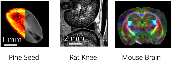
The magnetic field strength of the magnet is 11.7 T. With its high field strength the device can be utilized in anatomical imaging and quantitative analysis of unorthodox materials and samples (MRI-wise) . Furthermore, the high field strength provides an improved signal-to-noise ratio, and thus reduced scan times. Samples typically considered poorly visible in MRI may also be imaged thanks to the high quality hardware and imaging protocols as well as the high magnetic field strength. MicroMRI can microscopically image samples with below 30 µm resolution safely and non-invasively.
The diversity of the device is also emphasized with various RF coils that provide suitable approaches for each application. Typically 1H (i.e. protons, from for example water) are investigated and the MicroMRI device incorporates 5mm and 10mm (diameter) RF coils that provide the best quality for suitable samples. Also an 8mm 13C (carbon-13 isotope) RF coil can be used, providing fascinating and novel approaches and methods for the detection of carbon-induced signal.
General information and properties of the device are listed below.
BRUKER ULTRASHIELD 500 MHZ |
||
Field strength |
11.7T | 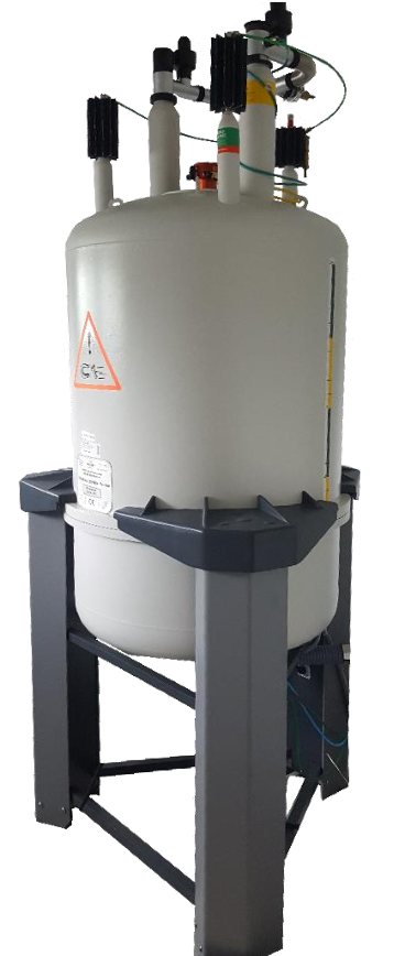 |
Image area |
10mm × 15mm (diameter × height) | |
Sample size |
< 10 mm (diam.). Height is not restricted but the region to be imaged must be at the bottom of the sample tube. | |
Resolution |
20µm | |
Scan time |
Seconds to hours. Depends on the resolution and what is needed from the sample. For a biological sample with a quantitative 3D MGE imaging sequence with approx. 35µm (isotropic) pixel size the imaging time is approx. 1h 50min. | |
RF coils |
5mm (1H) |10 mm (1H) | 8mm (13C) | |
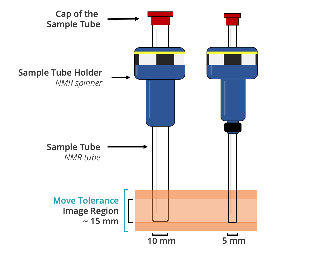
Measurement setting
The sample must fit at the bottom of a ⌀10mm sample tube. The image are is 15 mm in height but the image region can be moved 5-10mm (up/down, “move tolerance”). For smaller samples, a narrower sample tube can be used (⌀5mm) and a smaller RF coil providing a better resolution and signal-to-noise ratio.