Kanishka: How do we see stimulus-response in brain?
“Between stimulus and response there is a space. In that space is our power to choose our response. In our response lies our growth and our freedom.”
Have you ever jumped at a sudden loud noise or reflexively pulled your hand away from something hot? These quick, seemingly automatic reactions are your brain’s way of keeping you safe, showcasing the incredible speed and precision of the stimulus-response system. Every day, our brains handle a nonstop stream of stimuli—from the blare of an alarm clock to the warmth of a cup of coffee—each one triggering a carefully coordinated response.
But what’s really happening behind the scenes? How does your brain process these signals so rapidly, and why does it matter? In this blog, we will see what happens in the brain when it receives a stimuli, and how researchers practically measure those responses, in case of severe problems like Alziemer’s Disease or Epilepsy.

A plant’s leaves and stem move towards a light source. For the plant, light is the stimulus that triggers something (hormones/chemicals) inside the leaves to respond to the stimulus. This changes the plant’s direction towards (or away) from it.
Humans respond to stimuli in their own ways, depending on the kind of stimulus. But what happens in the brain during this stimulus-response process?
Our brain (and spinal cord, and nerves from the spinal cord, and gut, and…) is made up of specialized cells called neurons. These are specialized cells because communication between neurons is tightly regulated and faster compared to other cells of the body. Much faster (order of milliseconds!) than what it takes to get a meme viral.
You might have seen the word “brain activity” quite a lot, but what does it mean in principle? It is the summed-up activity of a group of neurons. These are called Field Potentials, as what we measure is the voltage deflection from the currents generated through neurons.
A neuron sends the message to other neurons in the form of chemicals and these travel to other neurons. This either decreases or increases the number of ions (inside and outside the neuron), and thus creates a dipole. Electrodes detect the sum of positive and negative charges in their vicinity. A single neuron’s dipole is too small to be measured as far away as the scalp. However, because electrodes detect the sum of charges in their vicinity, the dipoles from multiple neurons in a region will sum together. In the brain, cognitive processes require synchronized activation of a large groups of neurons. So, when the neurons receive a stimuli, they respond together and their potentials sum up and create a stronger, detectable electrical field.
EEG vs LFP?
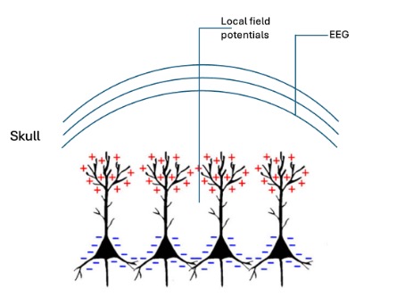
This activity could be recorded either at a global scale, i.e. from the scalp, or from the cortex called EEG, Electroencephalogram, or even locally from a particular area of the brain. The latter is thus called local field potential (LFPs) since these arise from the synchronised activity of a very specific area, and so, better spatial resolution compared to scalp EEG.
LFPs in “Hippocampus” are particularly important since the (ab)normal patterns of neural activity in this area is linked to behaviours like sleep and memory.
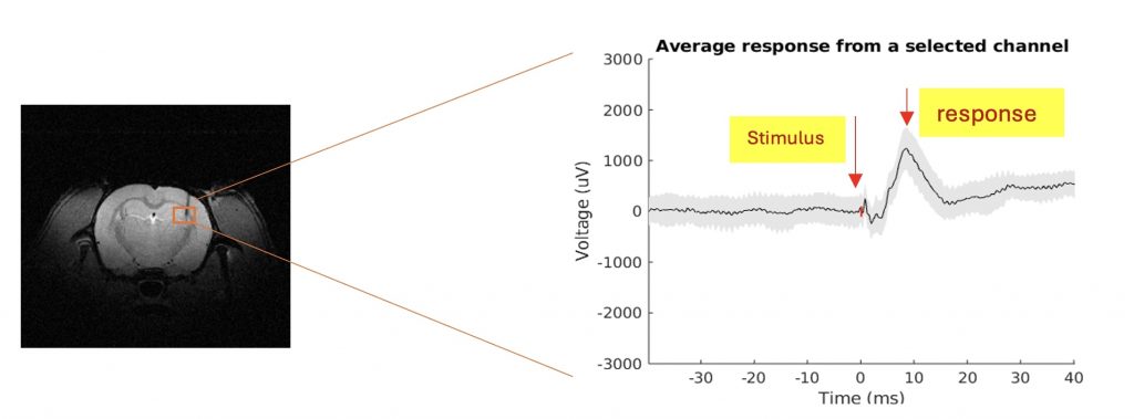
Thus, Local Field Potentials (in rat brain) could be used to study short-timed events (like unpredictable spread of epileptic activity), yielding good temporal and spatial resolution, but only for a minimum volume.
So, to cover the whole brain, we need another modality. Let’s see in the next blog how can we track stimulus-response events in the whole brain!
References
Jackson AF, Bolger DJ. The neurophysiological bases of EEG and EEG measurement: a review for the rest of us. Psychophysiology. 2014 Nov;51(11):1061-71. doi: 10.1111/psyp.12283. Epub 2014 Jul 17. PMID: 25039563.
Trueman, Shanon. “Phototropism Explained.” ThoughtCo, Apr. 5, 2023, thoughtco.com/phototropism-419215.
Kanishka works as a doctoral researcher in the Neuro-Innovation PhD Programme. Her research focuses on functional imaging of brain-wide networks associated with momentary events.