Melina Estela Dalmau: Hippocampus: a structure to remember
I’ve been exploring (and getting lost in) the microscopic world of brain tissue for almost two years now. The reason is that part of my PhD has to do with high-magnification images (aka photomicrographs) of rat brains. Honestly, I’m still amazed every time I zoom in on these images, discovering all the patterns and the complex arrangement of brain cells. But there is one particular brain region that caught my eye, not only for its pattern diversity but also for its role.
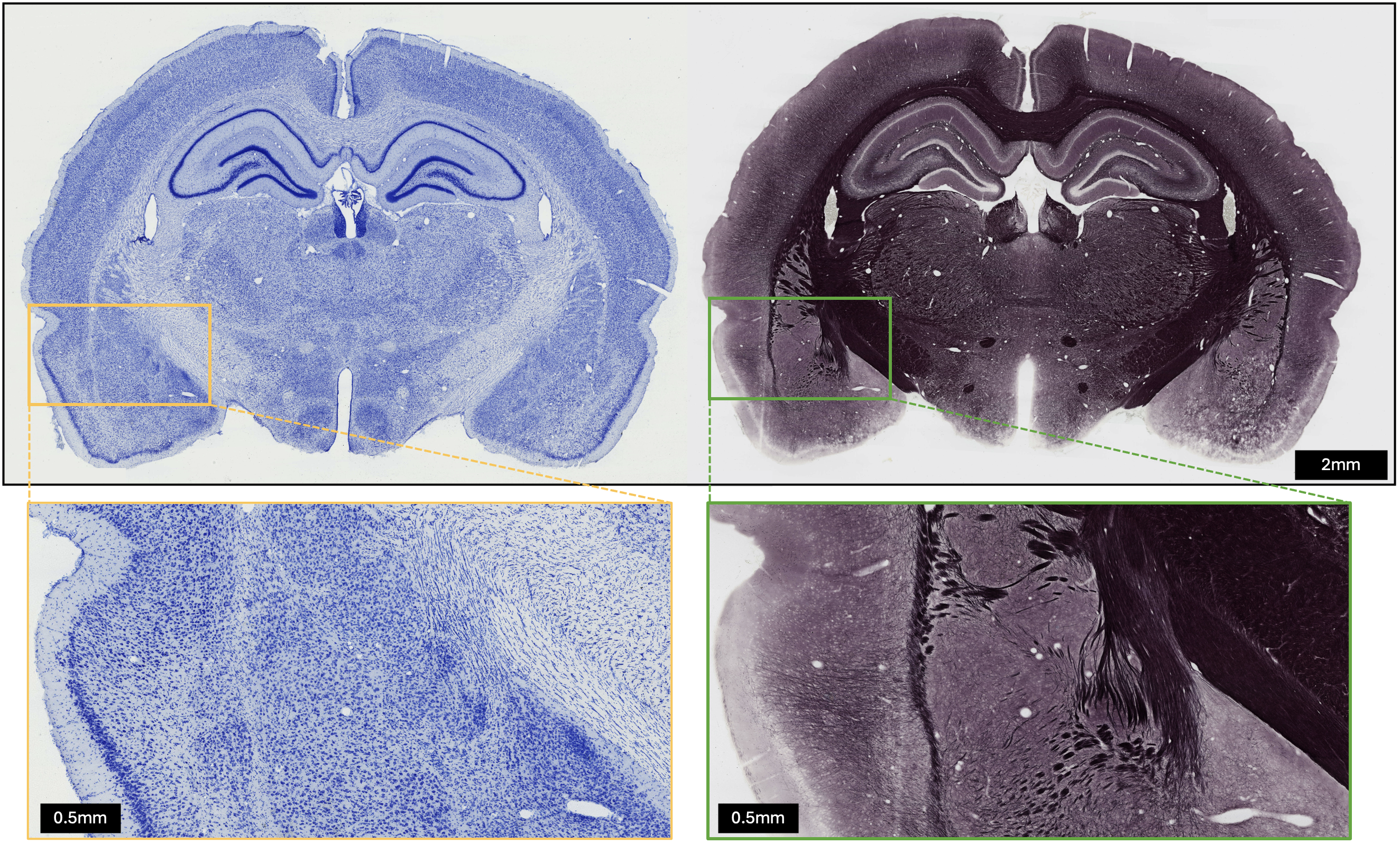
Let’s start by looking at the whole picture of a rat brain section. The image below contains two consecutive coronal sections that have been stained to enhance different cellular components. The left section shows cell bodies in blue, so we can observe how cells distribute, and how they differ in size and shape (more visible in the zoom-in view). The right section intensity denotes the presence of myelin, an insulating cover of most axons in the brain. With this stained section we can infer how myelinated axons are arranged throughout the tissue.

Interestingly, the organization of the brain tissue components is far from random; they are arranged in layers and groups that specialize in different functions. This organization appears to be key, as other species have larger brains than ours but do not necessarily show higher intelligence or cognitive complexity.
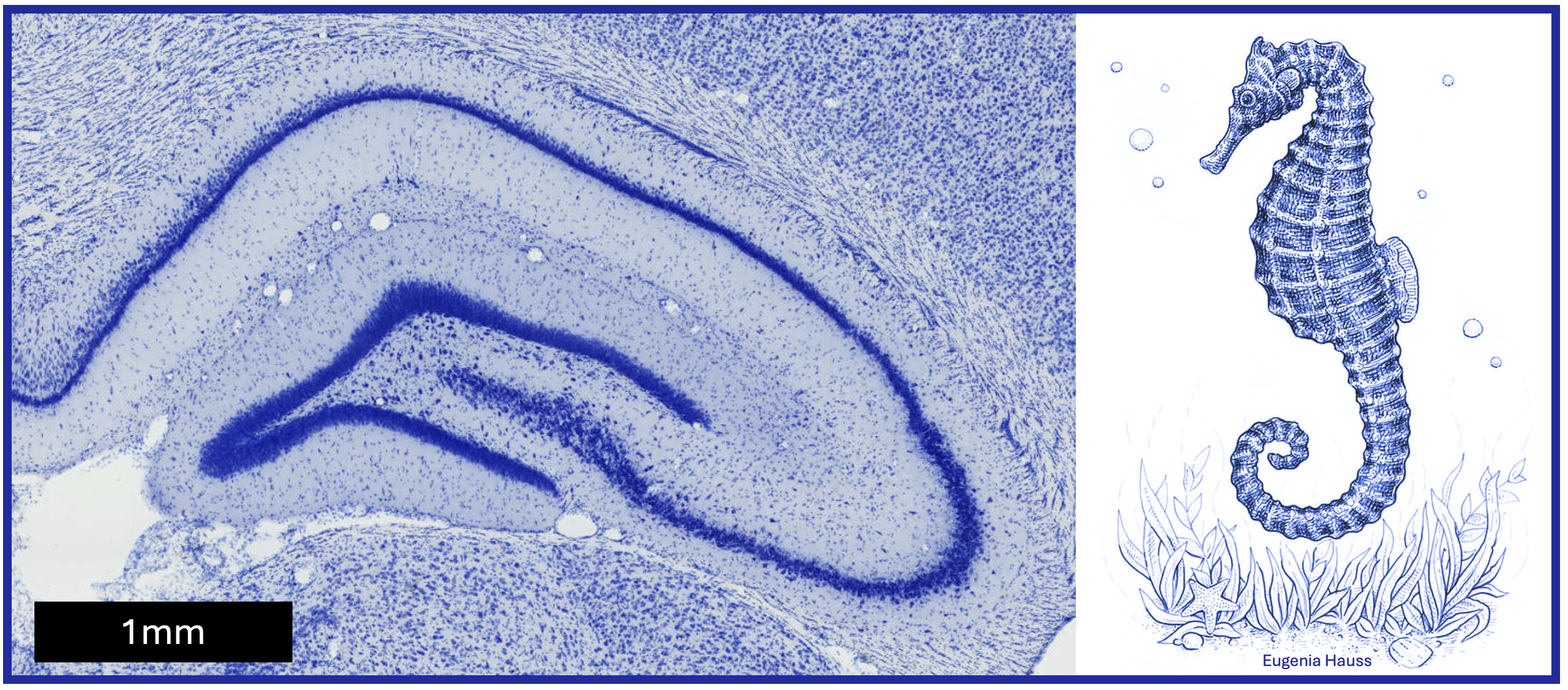
This is where the hippocampus comes in: a small, curved brain region in each hemisphere with a distinct (and beautiful) layered organization.
First, why call a brain region hippocampus? The term is actually another word for seahorse, derived from the Greek words hippos (horse) and kampos (sea monster). With that in mind, it’s easier to imagine how this curved brain region got its name —though spotting the seahorse shape in the 2D rat brain section might take a bit of creativity. You can test your skills with the following picture!

But what about hippocampus’ function? Over the years, different theories have been developed, but its role in memory formation, storage, and spatial navigation is now well-established.
Defining the functions of brain regions is not straightforward, and how the hippocampus became linked to memory formation was through the case of a person with epilepsy.
In 1953, as a treatment for his severe epileptic seizures, Henry Molaison underwent a surgical resection of most of his hippocampi. The surgery helped to reduce the seizures, but unexpectedly Henry was unable to create new memories (a condition called anterograde amnesia). That is, no chance of remembering anything new at all. Can you imagine living stuck in your current state of knowledge and being? Heavy, but that’s how we now know that the hippocampus is one of the few regions in the brain where new neurons are generated in adulthood (aka neurogenesis). And this formation of new neurons along with the strengthening of existing connections is key for learning and retaining.
Luckily, not all hippocampus stories are as tragic as Henry’s. Another example involves London taxi drivers. Studies have shown that their posterior hippocampi are significantly larger than those of control subjects. This example, complemented by other studies, established the relationship between hippocampus and navigation skills, hippocampus being crucial for processing spatial information and creating mental maps of the environment.
But of course, age and disease also play a role. In Alzheimer’s, the hippocampus is one of the first regions to deteriorate, leading to memory loss and also disorientation (the two attributes we have just described). Besides, traumatic brain injuries (i.e., whatever head impact) can also affect the hippocampus, causing issues with forming new memories. Here the bilateral symmetry of the brain protects us to some extent, since we have two hippocampi, one in each hemisphere. So, if the damage occurs in one side, the brain can retain nearly-normal memory functioning! In any case, whether healthy, diseased or by accident, old memories often remain intact, leading to the hypothesis that over time, the hippocampus transfers memories to other regions of the brain for long-term storage.
These are just some of the functions attributed to the hippocampus, but many questions remain open. We know its role in memory formation, but we still don’t know how it encodes, stores, and retrieves memories. We know it can grow when there is a need to retain information from the environment, but the exact mechanism by which hippocampal neurons create mental maps is unclear. We also know that hippocampus is highly vulnerable to damage and aging, but can we prevent those with cognitive training or physical activity?
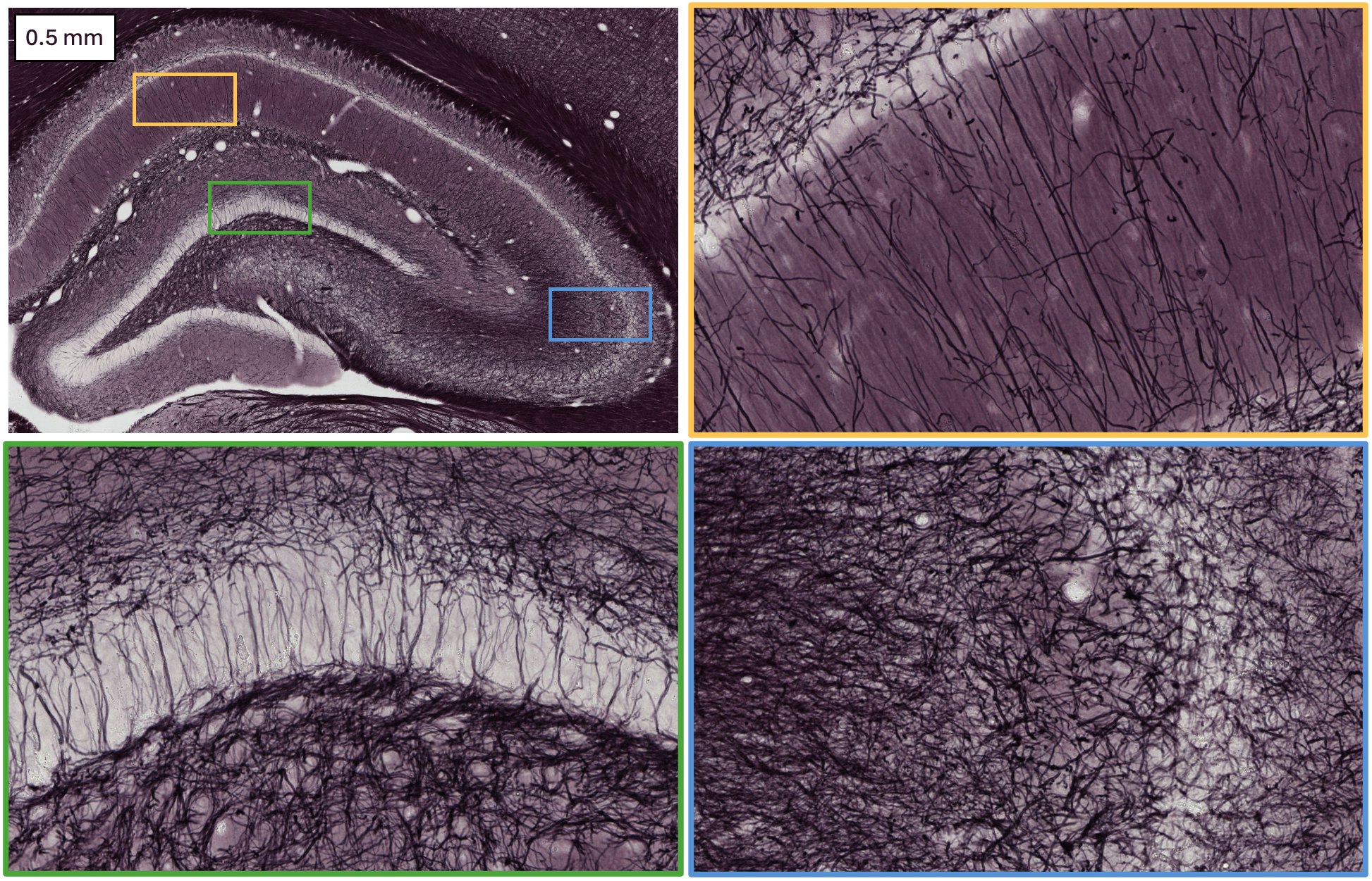
I hope now you understand better my fascination for this brain region. If not yet, here a last picture of the hippocampus:

Isn’t it crazy that those myelinated axons, ordered in layers, might help you remember what you just read?
References
The three-dimensional organization of the hippocampal formation: A review of anatomical dat. Amaral & Witter; Neuroscience, 1989.
The remarkable, yet not extraordinary, human brain as a scaled-up primate brain and its associated cost. Herculano-Houzel; PNAS, 2012.
The Legacy of Henry Molaison (1926–2008) and the Impact of His Bilateral Mesial Temporal Lobe Surgery on the Study of Human Memory. Dossani, Missios & Nanda; World Neurosurgery, 2015.
Navigation-related structural change in the hippocamapi of taxi driver. Maguire et al; PNAS, 2000.
Engrams and circuits crucial for systems consolidation of a memory. Kitamura et al; Science, 2017.
Melina Estela Dalmau works as a doctoral researcher in the Neuro-Innovation PhD programme. Her research seeks to bridge the gap between imaging and brain tissue.