Melina Estela Dalmau: Mielina
Do you remember the first time you learned we have neurons in the brain?
I have quite a strong memory from that time… but the reason why has less to do with science, and more to do with myself. Let me explain. I was in school when the teacher explained that a neuron is a special type of cell with three main parts: the soma (the cell body), the dendrites (which receives signals) and the axon (which sends them). This unique morphology makes neurons built for one thing above all else: communication. Neurons fire electrical signals that sweep along the axon like a wave. When the signal reaches the axon terminals it triggers the release of chemical messengers that pass on info to the next neuron, or to other cells like muscle or gland cells.
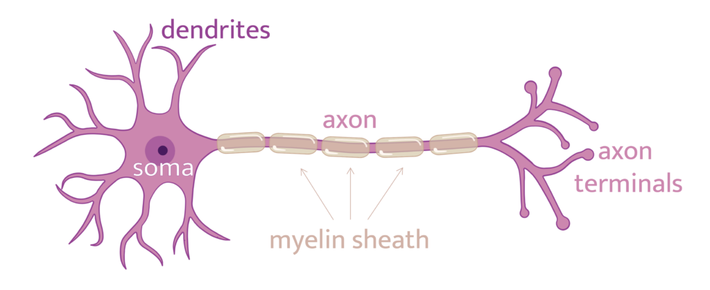
Importantly, most axons in the brain are wrapped in an insulating layer called myelin, which makes those electrical signals travel much faster. Actually, up to 100 times faster.
Why do I have such a strong memory of that class? Because in Spanish, myelin is mielina, and my name being Melina… You can imagine that many kids started to call me Mielina for a while.
Ironically, what I would have never guessed is that twenty years later, my PhD would be about myelin. And in this blog, I want to share some things I’ve learned since then.
Myelin is not a part of a neuron, but another cell’s full-time job
It could make sense that neurons made their own insulation (myelin sheaths), but… They don’t. Myelin sheaths come from a completely different cells called oligodendrocytes.
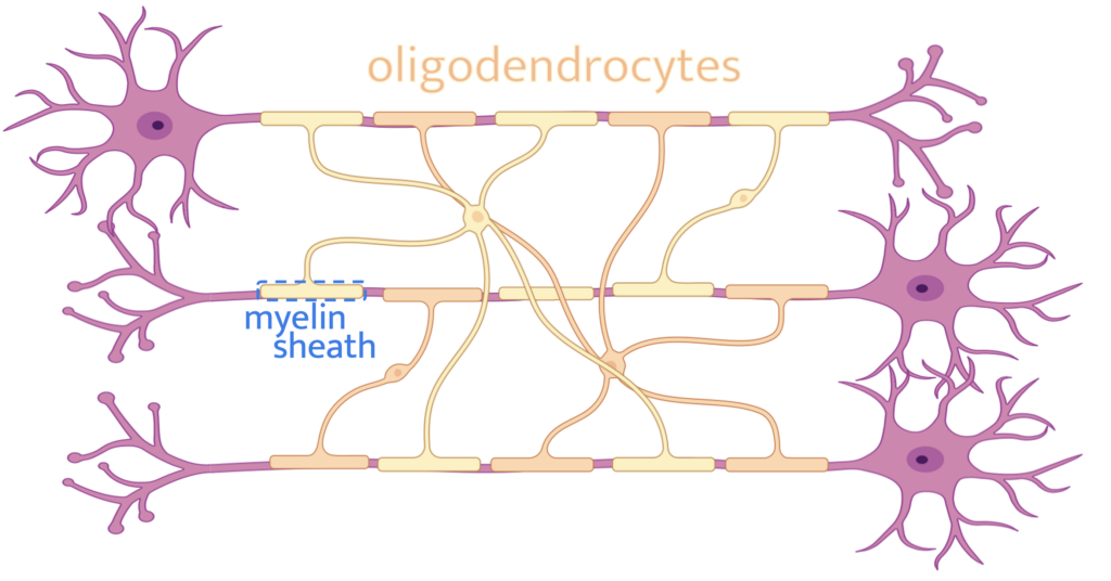
The name oligodendrocyte is itself a bit ironic. It literally means “cell with few branches” (in Greek: oligo = few, dendro = branch) because when the cell was first observed with rudimentary staining techniques in 1921, it appeared only to have a few stubby projections. But with modern technology, we’ve found quite the opposite. I like to imagine oligodendrocytes as an octopus (in reality, 40-70 pus!). Each of its tentacles reaches out and wraps itself around nearby axons in tight, concentric layers (the myelin sheath). Given the amount of tentacles, each oligodendrocyte may wrap several times the same axon as well as different ones. That is done with a safety rule: the same oligo won’t wrap the same axon twice in a row. So multiple oligos are needed to cover the whole axon of a neuron.
Myelin does not cover axons completely, the gaps are crucial
In the extreme case of an axon without any myelin, the electrical signal has to travel continuously along the whole axon-length, which is really slow. With the myelin sheaths (that remember, are insulating layers), the signal jumps from gap to gap, skipping the insulated regions entirely. This is called saltatory conduction, from the Latin saltare, to jump.
Let me make you feel myelin: you hit your toe. Without myelin, that info would take about a second to reach your brain. With myelin, the trip takes a hundredth of a second. And also, less energy is needed to do so. Hard to believe, but those 1μm gaps make all that difference.
Myelin damage: demyelination
But myelin isn’t just about speed. Damaged myelin means signals slow down, but the signals can also get scrambled, or even stop reaching their destination. Depending on where in the brain or spinal cord the damage occurs, demyelination can lead to blurred vision, muscle weakness, pain, difficulty with coordination, or cognitive impairment. One well-known disease caused by demyelination is multiple sclerosis (MS), in which the immune system mistakenly attacks myelin sheaths, resulting in damaged areas called lesions.
Interestingly, unlike neurons, oligodendrocytes can regenerate. And some remyelination happens spontaneously in MS lesions. But not preventing the development of the disease. Some questions researchers are trying to answer is why remyelination fails where it does, and how to help our oligo octopi along.
MS is the most studied case, but not the only one. Emerging research is connecting myelin disruption to schizophrenia, depression, and aspects of autism. Historically, these conditions have been studied from a purely neurological or psychological angle, without considering myelin’s role in connecting brain regions. We’ll see how this line of research develops.
Myelin is shaped by what you do
Myelin is not fixed. It changes throughout life based on what you do.
Learning to juggle literally increases myelination in the circuits responsible for tracking moving objects and coordinating your hands. And that’s true for any skill. Practice doesn’t just strengthen connections between neurons, it builds more myelin around circuits that are being repeatedly used.
And surprisingly, myelin can also be spent. A group of researchers scanned the brains of marathon runners before and after a race and found that myelin content dropped significantly in regions involved in motor coordination and sensory integration. But no worries, runners; it recovered within two months. One interpretation is that in extreme conditions, the brain may use myelin lipids as an energy reserve.
So, to recap:
Every action you do regularly will modify your oligodendrocytes, making them wrap the relevant circuits in more myelin. This will allow information going both to and from your brain to jump across your body much faster. And if the myelin gets damaged or used up, it’ll even regenerate!
Fast, adaptable and resilient. Turns out that Mielina was a cool nickname after all.
References:
Sabatino, J.J., Cree, B.A.C. and Hauser, S.L. (2025). New Horizons for Multiple Sclerosis Therapy: 2025 and Beyond. Annals of Neurology, 98: 317–328.
Valdés-Tovar, M. et al. (2022). Insights into myelin dysfunction in schizophrenia and bipolar disorder. World Journal of Psychiatry, 12(2): 264–285.
Wang, Y., Xiong, Z., Chen, M. and Zhu, Z. (2026). Myelin damage in major depressive disorder. Neuroscience & Biobehavioral Reviews, 184: 106607.
Adès, N. and Bouslama-Oueghlani, L. (2026). Myelin dysfunction in autism spectrum disorder. Molecular Psychiatry.
Ramos-Cabrer, P. et al. (2025). Reversible reduction in brain myelin content upon marathon running. Nature Metabolism, 7: 697–703.
Melina Estela Dalmau works as a doctoral researcher in the Neuro-Innovation PhD programme. Her research seeks to bridge the gap between imaging and brain tissue.