Facilities
Kuopio BIU equipment consist of five MRI scanners, including one with simultaneous PET/MRI, μCT and μPET unit. Unit can also provide access to optical, ultrasound and photo acoustic small animal imaging situated within the university. For MRI most of the contemporary imaging and spectroscopy pulse sequences are available or can be implemented on request. μPET imaging using 18F and 11C labelled tracers is performed in collaboration with Kuopio University Hospital and its radiochemistry laboratory and cyclotron. Through this collaboration, PET imaging with tailor made radiopharmaceuticals can be attained.
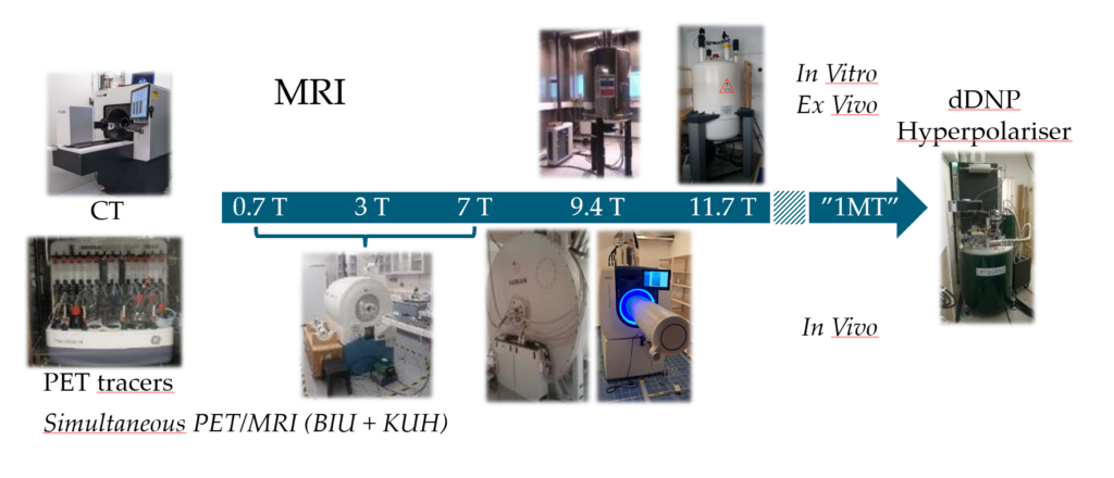
MRI
Kuopio Biomedical Imaging Unit houses five experimental MRI scanners operating from 0.7 T to 11.7T. Pre-clinical Hyperpolariser, SpinAligner (from The Technical University of Denmark), with adjustable polarisation field 3.35-10.1 T is situated next to 9.4 T horizontal imaging magnet.
- 9.4 T/31 cm horizontal MRI magnet (Agilent DirectDrive, VNMRj 3.1 and OpenVNMRj, and Bruker Biospec, PV 5.1 and 6.01) with 1H and X channels : Animals up to rabbits
- 9.4 T/16 cm horizontal Bruker BioSpec (PV360) magnet with 1H: Animals up to rats
- Horizontal simultaneous PET/MRI system with variable field strength (0.7 T, 3 T or 7 T)/16 cm , 1H and X channels and 1H cryoprobe for 7 T(MR Solutions) : Animals up to rats
- Vertical 9.4 T/89 mm magnet (Agilent DirectDrive, VNMRj 3.1) : Animals up to mice, materials, cells, tissues
- Vertical 11.7 T/20 mm magnet (Bruker MicroImaing, PV 6.0.1) with 1H and X channels : Materials, cells and tissues
PET
Pre-clinical PET/MRI system allows simultaneous imaging of one rat or up to three mice. System also has variable field (0.7 T, 3 T or 7 T). It is mainly used for imaging with various biomolecules labeled with e.g. 18F, 11C or 89Zr.
µCT
The MILabs U-CTUHR provides high-resolution in vivo anatomical imaging. The unit is suitable for imaging mice and rats. The CT unit has 7 µm focal spot size, maximum transaxial field-of-view of 13.5 cm and resolution of down to 15µm in focus mode. The voltage range of X-Ray tube is 10-65 kV.