Microglia in α-synucleinopathies
Microglia phagocytose α-synuclein aggregates which leads to release of proinflammatory cytokines, and in contrast, we have shown that presence of these cytokines may inhibit phagocytosis of aberrant proteins by human microglia (Niskanen et al., 2024). This causes inflammatory damage to neighbouring cells, leading to fewer microglia being able to take up and degrade α-synuclein, resulting in a cycle of neuroinflammation and protein aggregation that may cause further dysfunction and damage to surrounding cells.
Human microglia have differential gene expression compared to mouse, including higher expression of Parkinson’s-related genes, therefore studies carried out in rodent models alone do not translate to humans due to differences in microglial biology. Further, these highly reactive cells behave differently in vitro compared to in vivo, meaning that in vitro findings may not always translate to the in vivo situation. To address this, we established a novel model: transplantation of human microglia progenitors from induced pluripotent stem cells to an immunodeficient mouse model of Parkinson’s disease (Albert et al., 2025). We observed a human-specific effect of microglia on the α-synuclein aggregates, resulting in neuroprotection in the model. We now aim to elucidate this further.
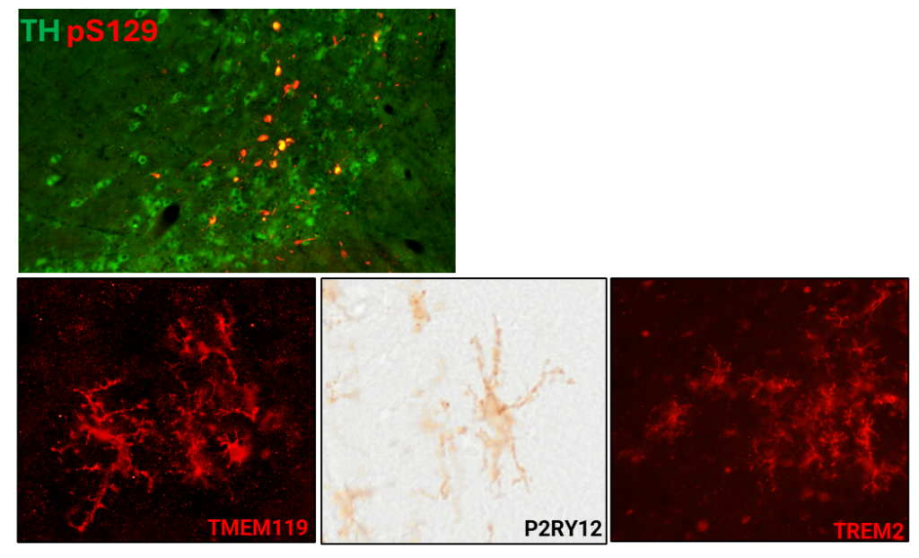
Top: Immunohistochemical staining of the mouse substantia nigra area with alpha-synuclein phosphorylated at serine-129 in the tyrosine hydroxylase cell bodies and surroundings. Bottom: Human microglia progenitors derived from induced pluripotent stem cells were transplanted to the mouse brain and 3 months later show a mature, brain-resident phenotype at the protein level.
Publications
Albert, K., Peltonen, S., Vanne, A., Kälvälä, S., Syvänen, V., Koistinaho, J., Luk, K.C., Lehtonen, Š., 2025. Human microglia reduce alpha-synuclein aggregation and are neuroprotective in adult mouse brain. Brain, Behavior, and Immunity 130, 106097. https://doi.org/10.1016/j.bbi.2025.106097
Niskanen, J., Peltonen, S., Ohtonen, S., Fazaludeen, M.F., Luk, K.C., Giudice, L., Koistinaho, J., Malm, T., Goldsteins, G., Albert, K., & Lehtonen, Š. (2024) Uptake of alpha-synuclein preformed fibrils is suppressed by inflammation and induces an aberrant phenotype in human microglia. Glia. https://doi.org/10.1002/glia.24626
Albert, K., Goldsteins, G., Kälvälä, S., Jolkkonen, J., & Lehtonen, Š. (2024) Glial cell transplant for brain diseases: the supportive saviours? Translational Medicine Communications, 9, 22. https://doi.org/10.1186/s41231-024-00182-y
Albert, K., Kälvälä, S., Hakosalo, V., Syvänen, V., Krupa, P., Niskanen, J., Peltonen, S., Sonninen, T.-M., & Lehtonen, Š. (2022) Cellular Models of Alpha-Synuclein Aggregation: What Have We Learned and Implications for Future Study. Biomedicines, 10, 2649. https://doi.org/10.3390/biomedicines10102649
Albert, K., Niskanen, J., Kälvälä, S., & Lehtonen, Š. (2021) Utilising Induced Pluripotent Stem Cells in Neurodegenerative Disease Research: Focus on Glia. Int J Mol Sci, 22. https://doi.org/10.3390/ijms22094334