Lewy body-like inclusions are modelled routinely using either by a overexpression of α-synuclein (αSyn) coding SNCA or by inducing pre-formed fibrills (PFFs) of short length alpha-synuclein fibrils (1). Previously it has been shown that, while SNCA is a risk factor for causing Parkinson’s disease (PD) and it is an classical example for familial PD with αSyn aggregations, its expression levels, which could be controlled by the methylation status of the gene, might not to be increased in the sporadic PD cases (2), while controversy remains (3).
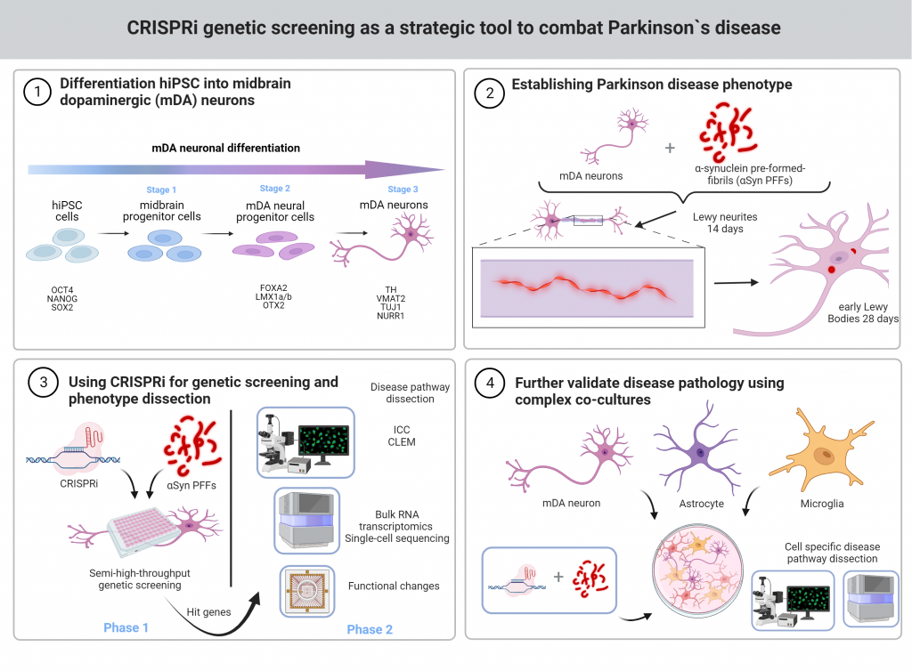
We aim to produce more authentic Lewy body-like inclusions by cultivating for longer periods of time after inducing with αSyn PFFs and in the correct conditions. We aim to see multicellular fractions as reported in PD (4). We aim to produce robust Lewy body pathology without the SNCA overexpression in a correct cultivation method, similar to Tanudjojo et al. (5). For this model we will conduct an accurate genetic screening and further dissect the disease pathway in mono- and co-cultures.
- Luk, K. C. et al. (2009) ‘Exogenous α-synuclein fibrils seed the formation of Lewy body-like intracellular inclusions in cultured cells’, Proceedings of the National Academy of Sciences of the United States of America, 106(47), pp. 20051–20056. doi: 10.1073/pnas.0908005106.
- Guhathakurta, S. et al. (2017) ‘Hypomethylation of intron1 of α-synuclein gene does not correlate with Parkinson’s disease’, Molecular Brain, 10(1), p. 6. doi: 10.1186/s13041-017-0285-z.
- Jowaed, A. et al. (2010) ‘Methylation Regulates Alpha-Synuclein Expression and Is Decreased in Parkinson’s Disease Patients’ Brains’, Journal of Neuroscience, 30(18), pp. 6355–6359. doi: 10.1523/JNEUROSCI.6119-09.2010.
- Shahmoradian, S. H. et al. (2019) ‘Lewy pathology in Parkinson’s disease consists of crowded organelles and lipid membranes’, Nature Neuroscience, 22(7), pp. 1099–1109. doi: 10.1038/s41593-019-0423-2.
- Tanudjojo, B. et al. (2021) ‘Phenotypic manifestation of α-synuclein strains derived from Parkinson’s disease and multiple system atrophy in human dopaminergic neurons’, Nature Communications, 12(1), p. 3817. doi: 10.1038/s41467-021-23682-z.