Equipment
Microscopes
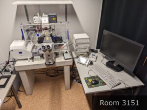
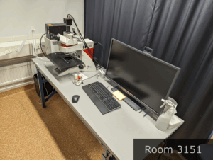
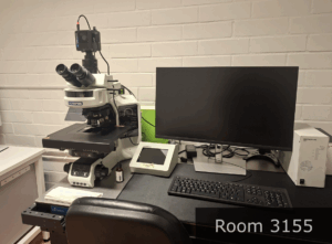
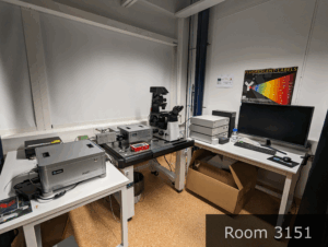
Other equipment
RESERVATIONS
Starting March 2026 all microscopes will be booked through OpenIris.
Reservation of Opera Phenix Plus, Harmony analysis computer and ISS M612 systems is done on OpenIris reservation system:
https://openiris.io/landing/?ReturnUrl=%2f#!/browse/resources
Here you find tutorials on how to book microscopes through OpenIris