EDUCATIONAL MATERIAL (NENS)
NENS Cluster videos on glia and inflammation
FENS is the voice of European neuroscience 21,000 neuroscientists, 44 member societies, 33 European countries. Click here for more information
Astrocytes and microglia in neuroinflammation
Glia and inflammation in Amyotrophic lateral sclerosis. Astrogliosis. Microglial phenotypes. Interaction of microglia and astrocytes with neurons. BBB breach and the role of astrocytes. Infiltration of immune cells in brain tissue and interaction with glia. Humoral factors of inflammation and glia. Comparative glial cellular morphology obtained by high resolution microscopy. Oxidative stress and microglia – release of cytokines.
Dynamical measurements from neuroglia in vitro
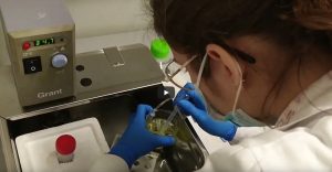
Preparation of primary rat microglia (and/or BV-2 cell line) /astrocytes for experiment. Dye loading – Ca2+– or ROS-sensitive dyes. Videomicroscopy setup. Recording software environment. Dynamical measurements of Ca2+ or ROS – transients. Signal analysis.
Here we show a protocol for recording the electrical activity organoids on MEA. The premise is the comparison of organoids with and without microglia (Fagerlund and Dougalis et al., 2021). We go through the main steps in slicing an organoid, preparing it for recording and doing baseline recording and pharmacological stimulations.
Protocol description starts at 2:10
Video edited by:
Anssi Pelkonen
Camera:
Ilkka Fagerlund
MEA experiment:
Mireia Gomez-Budia
Special thanks:
Neuroinflammation research group lead by Tarja Malm
Publication:
Fagerlund, I.; Dougalis, A.; Shakirzyanova, A.; Gómez-Budia, M.; Pelkonen, A.; Konttinen, H.; Ohtonen, S.; Fazaludeen, M.F.; Koskuvi, M.; Kuusisto, J.; Hernández, D.; Pebay, A.; Koistinaho, J.; Rauramaa, T.; Lehtonen, Š.; Korhonen, P.; Malm, T. Microglia-like Cells Promote Neuronal Functions in Cerebral Organoids. Cells 2022, 11, 124. https://doi.org/10.3390/cells11010124
Some images also from:
Pelkonen, A.; Pistono, C.; Klecki, P.; Gómez-Budia, M.; Dougalis, A.; Konttinen, H.; Stanová, I.; Fagerlund, I.; Leinonen, V.; Korhonen, P.; Malm, T. Functional Characterization of Human Pluripotent Stem Cell-Derived Models of the Brain with Microelectrode Arrays. Cells 2022, 11, 106. https://doi.org/10.3390/cells11010106
Sources otherwise indicated in the video
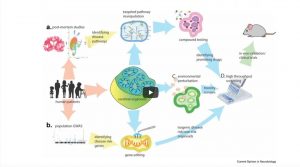
Modeling Neuroinflammation in human context
This lecture covers current knowledge on human-based models of neuroinflammation. These include iPSC-derived 2D and 3D models, possible differences between the models and their suitability for functional studies.
How to culture human iPSC-derived microglia
We show the detailed protocol and practical tips for differentiation of microglia-like cells from induced pluripotent stem cells. We will introduce methodology to carry out quality control analysis of microglia throughout the differentiation protocol using flow cytometry and how to carry out functional studies for phagocytosis and migration using Incucyte Live Imaging.
Here we describe how to differentiate human microglia-like cells from induced pluripotent stem cells (iPSC). This protocol is based on our publication investigating the impact of Alzheimer´disease -predisposing genetic backgrounds on functionality of human microglia-like cells (iMGLs) (Konttinen H., et al., 2019). In this video, we will go through the main steps of the protocol and guide you towards succesfull differentation. By following this protocol, large number of human microglia-like cells can be procuded to match you research needs!
Protocol description start at 2:20
Video produced and edited by:
Videopalvelu Merja Palm
Funded by:
Network of European Neuroscience Schools (NENS)
University of Eastern Finland, the Faculty of Health Sciences
Special thanks:
Neuroinflammation research group lead by Tarja Malm
Molecular Neurodegeneration group lead by Annakaisa Haapasalo
Part of the work was carried out with the support of UEF Cell and Tissue Imaging Unit, University of Eastern Finland, Biocenter Kuopio and Biocenter Finland.
Publication:
Konttinen H, Cabral-da-Silva MEC, Ohtonen S, Wojciechowski S, Shakirzyanova A, Caligola S, Giugno R, Ishchenko Y, Hernández D, Fazaludeen MF, Eamen S, Budia MG, Fagerlund I, Scoyni F, Korhonen P, Huber N, Haapasalo A, Hewitt AW, Vickers J, Smith GC, Oksanen M, Graff C, Kanninen KM, Lehtonen S, Propson N, Schwartz MP, Pébay A, Koistinaho J, Ooi L, Malm T. PSEN1ΔE9, APPswe, and APOE4 Confer Disparate Phenotypes in Human iPSC-Derived Microglia. Stem Cell Reports. 2019 Oct 8;13(4):669-683. doi: 10.1016/j.stemcr.2019.08.004. Epub 2019 Sep 12. PMID: 31522977; PMCID: PMC6829767.