Computed Tomography (CT)
Basic principles
Computed tomography (CT) is a medical imaging technique developed almost 40 years ago. Method uses digital geometry processing to generate a three-dimensional image from a large set of two-dimensional X-ray images taken around a single axis of rotation. The basic physics is similar to conventional X-ray imaging; different materials have different attenuation coefficients that are dependent on density of the material. In order to acquire good quality images, the sample has to have enough internal density variation. CT is used also in fields other than healthcare, for example nondestructive material testing.
University of Eastern Finland Kuopio campus area provides a continuum of computed tomography devices, beginning from micrometer-scale pre-clinical scanner to in vivo CT and further to clinical cone beam CT (Verity), pQCT and total body instruments.
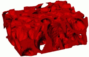
Micro-CT image stacks can be used as a basis of 3D-models. Meshes created from cross-sectional images can be used, for example, in acoustic and mechanical finite element modeling.
Specific aims
In our group computed tomography is currently being used for bone and articular cartilage research and for material characterization. Bone mineral density and structural parameters are often used as a reference technique when developing new methods for bone quality assessment. In articular cartilage research we aim to quantify cartilage quality and proteoglycan (PG) content, and to detect cartilage injuries.
Bone research
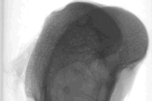
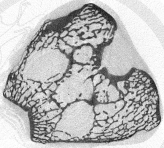
X-ray techniques have been used to image skeletal system since the discovery of X-rays. Computed tomography is no exception; it can be used to measure, for example, bone thickness, area and mineral density. With high-resolution scanners, also cancellous bone and even individual trabeculae are easily quantified. Structural data acquired with computed tomography is often used as a reference technique for morphological analyses.
Bone mineral density, as measured with dual-energy X-ray absorptiometry (DXA) and structural properties determined with micro-CT have been used as reference parameters for bone ultrasound studies.
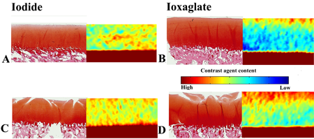
Cartilage research
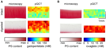
X-ray images, especifically the narrowing of knee joint space, have long been used in diagnosis of osteoarthrosis (OA). This method only shows changes when the cartilage has been destroyed and the damage is already irreversible. Recently, however, CT method (contrast enhanced computed tomography, CECT) analogous to the dGEMRIC has been used to quantify proteoglycan distribution and cartilage quality in articular cartilage [1-4]. These methods utilize ionic contrast agents and hypothesis that the negatively charged glycosaminoglycans (GAG) in proteoglycans cause a fixed charge density (FCD) variation in cartilage; ionic contrast agent is supposed to distribute relatively to the FCD. This method has potential for early diagnosis of OA, when the cartilage still looks visually healthy.
References
- Palmer AW, Guldberg RE, Levenston ME. Analysis of cartilage matrix fixed charge density and three-dimensional morphology via contrast-enhanced microcomputed tomography. Proc Natl Acad Sci USA 103: 19255-60, 2006.
- Cockman MD, Blanton CA, Chmielewski PA, Dong L, Dufresne TE, Hookfin EB, Karb MJ, Liu S, Wehmeyer KR. Quantitative imaging of proteoglycan in cartilage using a gadolinium probe and microCT. Osteoarthritis Cartilage 14: 210-4, 2006.
- Kallioniemi AS, Jurvelin JS, Nieminen MT, Lammi MJ, Töyräs J. Contrast agent enhanced pQCT of articular cartilage. Phys Med Biol 52: 1209-19, 2007.
- Silvast TS, Jurvelin JS, Aula AS, Lammi MJ, Töyräs J. Contrast agent-enhanced computed tomography of articular cartilage: association with tissue composition and properties. Acta Radiol 50:78-85, 2009.
- Kokkonen HT, Jurvelin JS, Tiitu V, Toyras J. Detection of mechanical injury of articular cartilage using contrast enhanced computed tomography. Osteoarthritis Cartilage 2011;19:295-301.
- Kokkonen HT, Aula AS, Kröger H, Suomalainen J-S, Lammentausta E, Mervaala E, et al. Contrast Enhanced Computed Tomography of Human Knee Cartilage in vivo. Cartilage 2012.
Ph.D. theses of our researchers
Antti Aula née Kallioniemi:
Publications of our researchers
- Kokkonen HT, Aula AS, Kröger H, Suomalainen J-S, Lammentausta E, Mervaala E, et al. Contrast Enhanced Computed Tomography of Human Knee Cartilage in vivo. Cartilage 2012.
- Kulmala K, Pulkkinen H, Rieppo L, Tiitu V, Kiviranta I, Brünott A, et al. Contrast-Enhanced Micro–Computed Tomography in Evaluation of Spontaneous Repair of Equine Cartilage. Cartilage 2012.