Ultrasonics
Basic techniques
The propagation of ultrasound depends on the mechanical properties of the material. For example in an elastic isotropic material the speed of sound in a given material depends on Young’s modulus, the density and Poisson’s ratio of the material, while the acoustic pressure is related to Young’s modulus and the particle displacement. When ultrasound propagates in biological tissues it undergoes attenuation, absorption, reflection, scattering and beam spreading. The ultrasound assessment of tissues is based on analyses of acoustical responses.
Quantitative ultrasound (QUS) parameters are usually measured in the through-transmission (TT) or pulse-echo (PE) geometries. The TT geometry requires the presence of two transducers on opposite sides of the object and this allows the quantification of the speed of sound and ultrasound attenuation in the object. One transducer transmits the ultrasound and the other records the signal transmitted through the object. TT measurements have been used in clinical bone densitometry. The potential of PE measurements has been growing recently. The pulse-echo geometry requires only one transducer and allows measurements of reflection and backscatter parameters. The ultrasound pulse is transmitted to the object and the returning signal is recorded with the same transducer. Quantitative ultrasound parameters are often normalized with reference measurements to minimize the system specific errors.
Ultrasonic assessment of articular cartilage
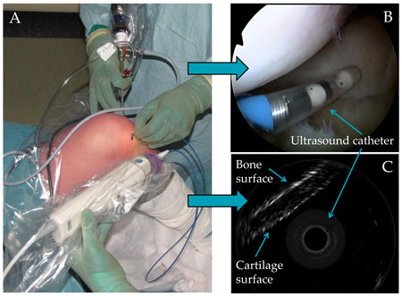
Osteoarthrosis is a severe cartilage disease, commonly diagnosed among elderly people. For a successful treatment of the disease, an early detection of the pathological changes in cartilage is essential, but present clinical applications provide foremost qualitative information of the tissue. The aim of ultrasonic assessment of articular cartilage is to reveal interrelationships between ultrasonic parameters and structural and mechanical properties of the tissue for the validation and development of quantitative clinical ultrasonic parameters and instruments.
Changes in the composition and structure of the articular cartilage related to the cartilage degradation or injuries have a significant effect on the acoustical properties of the tissue, thus, the integrity of the articular cartilage can be evaluated with quantitative ultrasound measurements. The literature describes various ultrasound methods for estimating the integrity of the articular cartilage. These techniques are based on measurements of the changes in the ultrasound speed, attenuation and backscattering within the cartilage and reflection coefficient and roughness of the articular surface.
Ultrasonic assessment of bone
Osteoporosis (OP) is a severe threat against the health of musculoskeletal system in elderly people, especially in women beyond the menopause. In OP the bone is resorpted diminishing the bone mineral density and thus leading to reduced bone strength and exposing bones to fractures. Early diagnosis of OP is of great importance for successful treatment and prevention of fractures.
Various parameters can be calculated from the measured signals, e.g., broadband ultrasound attenuation (BUA), speed of sound (SOS), integrated reflection coefficient (IRC) and broadband ultrasound backscattering (BUB), which have been shown to be related to bone micro-architecture.
The first aim of ours is to experimentally investigate the relationship between acoustic, structural, mechanical and compositional properties of trabecular and cortical bone. Second, to develop a novel instrument, which is currently under design and construction in our laboratory, for rapid multi-site evaluation of bone quality.