Microscopy
Polarized light microscopy
All fibrillar structures, including collagen fibrils, are birefringent objects, i.e. they are capable of altering the state of polarization. A quantitative technique for measuring birefringence of any transparent sample has been developed and utilized here in University of Eastern Finland (formerly known as University of Kuopio) [1-3]. The Polarized light microscope has two linear polarizers perpendicular to each and no light is passed through them, i.e. only dark field is seen without the specimen. An unstained sample is put between two linear crossed polarizers. If the sample is optically anisotropic, the state of polarization is altered and light passes through the second linear polarizer. Birefringence can be determined by measuring grayscale images, which are then converted to birefringence according to inverse-Fresnell equation.
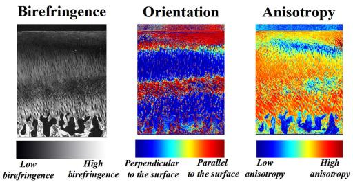
Quantitative polarized light microscopy is used in our laboratory to analyze collagen network of articular cartilage. We have recently developed an enhanced polarized light microscope (EPOL) [4], which is capable of measuring birefringence, orientation and anisotropy of any birefringent material suitable for microscopical investigation (Figure 1). Information from analysis is used to study different models of osteoarthritis (OA), biomechanics of articular cartilage, structure-function relationships, in phenotyping genetic animals and so on. The technique, however, can be used to study other biological tissues such as tendon, muscle, skin and bone or when information about polymers or other inorganic materials is required.
Digital densitometry
The amount of different substances can be measured optically based on color reactions between the molecule of interest and a specific dye. The dye molecule must have stoichiometric binding. i.e. a unambiguous relation between the dye molecule and measured molecule concentration must exist. Optical density measurement is based on the Beer-Lambert’s Law. The law states that the absorbance (optical density) of a substance is directly proportional to the concentration of that substance.
Digital densitometry can be utilized to measure proteoglycan (PG) concentration of safranin O-stained histological sections [5-6]. Measurements are conducted with a microscope using monochromatic light, and the images are captured with a CCD-camera. Calibration of the images is conducted using neutral density filters. The advantage of concentration analysis based on quantitative microscopy is that spatial distribution of dye, i.e. substrate that dye binds, can be measured pixel by pixel from microscopical sections. This approach gives much more information than conventional biochemical analysis of tissue extracts.
References
- Arokoski JP, Hyttinen MM, Lapveteläinen T, Takács P, Kosztáczky B, Módis L, Kovanen V, Helminen H. Decreased birefringence of the superficial zone collagen network in the canine knee (stifle) articular cartilage after long distance running training, detected by quantitative polarised light microscopy. Ann Rheum Dis. 1996 Apr;55(4):253-64.
- Király K, Hyttinen MM, Lapveteläinen T, Elo M, Kiviranta I, Dobai J, Módis L, Helminen HJ, Arokoski JP. Specimen preparation and quantification of collagen birefringence in unstained sections of articular cartilage using image analysis and polarizing light microscopy. Histochem J. 1997 Apr;29(4):317-27.
- Kocsis K, Hyttinen M, Helminen HJ, Aydelotte MB, Módis L. Combination of digital image analysis and polarization microscopy: theoretical considerations and experimental data. Microsc Res Tech. 1998 Dec 15;43(6):511-7.
- Rieppo J, Hallikainen J, Jurvelin JS, Kiviranta I, Helminen HJ, Hyttinen MM. Practical considerations in the use of polarized light microscopy in the analysis of the collagen network in articular cartilage. Microsc Res Tech. 2008 Apr;71(4):279-87.
- Kiviranta I, Jurvelin J, Tammi M, Säämänen AM, Helminen HJ. Microspectrophotometric quantitation of glycosaminoglycans in articular cartilage sections stained with Safranin O. Histochemistry. 1985;82(3):249-55.
- Király K, Lapveteläinen T, Arokoski J, Törrönen K, Módis L, Kiviranta I, Helminen HJ. Application of selected cationic dyes for the semiquantitative estimation of glycosaminoglycans in histological sections of articular cartilage by microspectrophotometry. Histochem J. 1996 Aug;28(8):577-90.