Optical Imaging
Detection, grading and delineation of articular cartilage degeneration and injuries from arthroscopic images have been considered inaccurate and poorly repeatable because the diagnosis is based on visual evaluation only [1, 2].
Optical coherence tomography (OCT) is a promising technique for detection, delineation and quantitative assessment of cartilage lesions [3]. It is an interferometer based optical imaging technique where reflected and backscattered near-infrared light from different depths is measured. Light reflection and scattering properties of a tissue are related to tissue structure and composition [4].
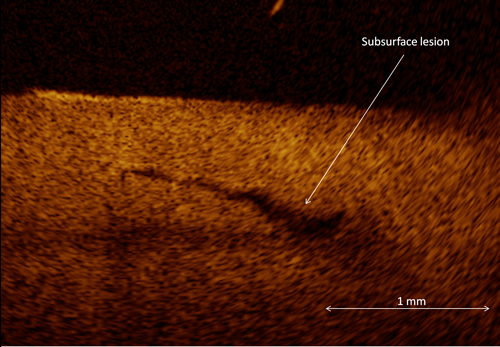
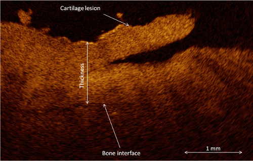
We study optical coherence tomography in articular cartilage imaging. OCT provides high spatial resolution (up to 10 µm) and is ideally suited for both visual assessment of cartilage lesions and quantitation of surface roughness [5, 6]. Unlike conventional arthroscopy technique, OCT also enables detection of subsurface lesions (Fig. 1) and measurement of cartilage thickness [7] (Fig. 2). Furthermore, quantitative reflection and backscattering analysis can be performed [5, 8]. Our aim is to study how articular cartilage degeneration and injuries affect the light reflection and scattering properties of the cartilage and how the state of the cartilage could be assessed repeatably and reliably from quantitative optical information. Miniaturized OCT probes enable arthroscopic OCT imaging in vivo and development of multimodality imaging techniques.
References
- Spahn, G., H.M. Klinger, and G.O. Hofmann, How valid is the arthroscopic diagnosis of cartilage lesions? Results of an opinion survey among highly experienced arthroscopic surgeons. Arch Orthop Trauma Surg, 2009. 129(8): p. 1117-21.
- Spahn, G., et al., Reliability in arthroscopic grading of cartilage lesions: results of a prospective blinded study for evaluation of inter-observer reliability. Arch Orthop Trauma Surg, 2011. 131(3): p. 377-81.
- Viren, T., et al., Comparison of ultrasound and optical coherence tomography techniques for evaluation of integrity of spontaneously repaired horse cartilage. J Med Eng Technol, 2012. 36(3): p. 185-92.
- Walther, J., et al., Optical coherence tomography in biomedical research. Anal Bioanal Chem, 2011. 400(9): p. 2721-43.
- Saarakkala, S., et al., Quantification of the optical surface reflection and surface roughness of articular cartilage using optical coherence tomography. Physics in Medicine and Biology, 2009. 54(22): p. 6837-6852.
- Chu, C.R., et al., Arthroscopic microscopy of articular cartilage using optical coherence tomography. American Journal of Sports Medicine, 2004. 32(3): p. 699-709.
- Han, C.W., et al., Analysis of rabbit articular cartilage repair after chondrocyte implantation using optical coherence tomography. Osteoarthritis Cartilage, 2003. 11(2): p. 111-21.
- Huang, Y.P., et al., Effects of optical beam angle on quantitative optical coherence tomography (OCT) in normal and surface degenerated bovine articular cartilage. Phys Med Biol, 2011. 56(2): p. 491-509.