The CT Group (Cartilage Theranostics & Computed Tomography)
Left (front to back): Janne Mäkelä, Jiri Jäntti, Petri Paakkari, Milka Poimala, Maria Rissanen, Juuso Tuppurainen
Right: Elizabeth George, Afifah Tsurayya, Heta Mertano
Principal investigators
Postdocs
Doctoral researchers
Collaborators
- Professor Brian Snyder – Boston Children’s Hospital
- Professor Mark Grinstaff – Boston University
- Assistant Professor Michael Albro – Boston University
- Professor Seppo Koskinen – Karolinska Institut
- Professor Miika Nieminen – Oulu University
- Professor Travis Klein – Queensland University of Technology
- Professor René van Weeren – Utrecht University
- Professor Jos Malda – Utrecht University
- Associate Professor Greet Kerckhofs – KU Leuven
- Associate Professor Kathryn Stok – University of Melbourne
Overview
The CT Group (Cartilage Theranostics & Computed Tomography) is committed to early therapeutics and diagnosis of osteoarthritis. Our focus is to study the capability of new (nano) contrast media to image articular cartilage and to develop advanced computed tomography techniques to track disease progression over time. In addition, we are exploring the use of synthetic supplements to reinforce and lubricate the body’s tribosystems. To gain a deeper understanding of joint lubrication and function, we integrate experimental measurements with computational modeling techniques.
Current projects
New Generation Spectral Computed Tomography and Nano Contrast Media for Osteoarthritis Diagnosis
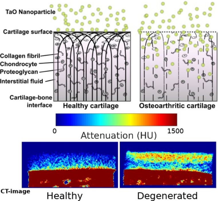
Spectral computed tomography represents the future of X-ray imaging (Fig. 1). Commercial instrumentation is not yet available in Finland. The technology allows retrospective detection of individual elements with only one scan, making it a superb modality to simultaneously trace multiple contrast agents. Concurrently, modern nanotechnology enables more effective design of active targeting contrast agents. The project develops a spectral computed tomography method for early diagnosis of articular cartilage injuries based on the simultaneous use of separate nano contrast agents. The solution to be developed will enable lower radiation doses in imaging, improved imaging accuracy, as well as new information for the optimization of future spectral imaging and nano contrast agents.
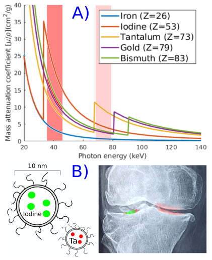
Fig. 1: A) Elements absorb X-ray attenuation characteristically (solid lines). Spectral-CT scanning uses energy bins (red bars) that allow separation of different element attenuation. B) Artistic illustration shows an example of imaginary CT image of a patient who has fallen off a bicycle. CT imaging is done using a Iodine and Tantalum NP contrast agent mixture. OA increases cartilage porosity to a point where properly sized nanoparticles diffuse inside the tissue. Bigger iodine NP accumulation reveals clear trauma, which will need reparative surgery. Smaller Tantalum (Ta) reveals mild idiopathic degeneration (fibrillation) at medial sites of both knees which can be treated using a reinforcing nanoviscosupplement.