Optical sensing and imaging
Spectral imaging
Keywords: spectral imaging; color; sensing applications for medicine, forestry, environment, and biology;
The group is a joint research group of the School of Computing and the Department of Physics and Mathematics. It belongs to the Center for Photonics Sciences. Group is also member of the Academy of Finland’s Flagship on Photonics Research and Innovation (PREIN).
Spectromics Laboratory is the first spectral imaging research environment in Finland focused in plant imaging.
Environmental and medical photonics
Keywords: biophotonics; biomedical imaging; neurophotonics; Raman spectroscopy; Surface-Enhanced Raman Spectroscopy (SERS) ; black silicon; gold nanoparticles; graphene; biocompatibility; enhancement factor; small organic molecules; living cells; nanodiamonds; color center; carbon nanotube; graphene quantum dot; theranostic agent
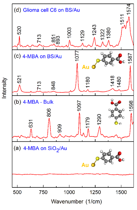
The group is currently focused on the design, modeling and fabrication of a range of surface-enhanced Raman spectroscopy substrates based on black silicon (bSi). We propose to sculpture the bSi surface enhanced with graphene and / or gold nanolayers in order to achieve SERS enhancement factor as high as 8 orders of magnitude. This finding makes the SERS-active bSi-based substrate suitable for a number of the environmental and / or biomedical applications when detecting analytes’ trace concentrations are required. Being biocompatible, the bSi/Au SERS-active substrate offers a unique opportunity to monitor the functional state of living cells using proteins / lipids / DNA/ RNA characteristic Raman bands as markers.
Highly uniform and reliable bSi based SERS substrates provide a pathway to the sensitive and selective, scalable, and low-cost lab-on-a-chip SERS biosensors that can be integrated into silicon-based photonics device.
In research, the group actively collaborates with international partners from Center for Physical Sciences and Technology, Vilnius, Lithuania.
Contact persons:
Prof. Polina Kuzhir
Prof. Yuri Svirko
Selected publications:
- L. Golubewa, R. Karpicz, I. Matulaitiene, A. Selskis, D. Rutkauskas, A. Pushkarchuk, T. Khlopina, D. Michels, D. Lyakhov, T. Kulahava, A. Shah, Y. Svirko, and P. Kuzhir, “Surface-enhanced Raman spectroscopy of organic molecules and living cells with gold-plated black silicon”, ACS Appl. Mater. Interfaces 12, 50971 (2020).
- L. Golubewa, H. Rehman, T. Kulahava, R. Karpicz, M. Baah, T. Kaplas, A. Shah, S. Malykhin, A. Obraztsov, D. Rutkauskas, M. Jankunec, I. Matulaitiene, A. Selskis, A. Denisov, Y. Svirko, and P. Kuzhir, “Macro-, micro- and nano-roughness of carbon-based interface with the brain cells: towards a versatile bio-sensing platform”, Sensors 20, 5028 (2020).
- L. Golubewa, et. al., “Visualizing hypochlorous acid production by human neutrophils with fluorescent graphene quantum dots”, Nanotechnology 33 095101 (2022).
- L. Golubewa, et. al., “Black Silicon: Breaking through the Everlasting Cost vs. Effectivity Trade-Off for SERS Substrates”, Materials 16, 1948 (2023).
- L. Golubewa, et. al., “Stable and Reusable Lace-like Black Silicon Nanostructures Coated with Nanometer-Thick Gold Films for SERS-Based Sensing”, ACS Applied Nano Materials 6, 4770 (2023).
Keywords:
Black silicon, surface-enhanced Raman spectroscopy, gold nanoparticles, graphene, biocompatibility, enhancement factor, small organic molecules, living cells

Comparison of Raman spectra of 4-MBA monolayer on SiO2/Au smooth substrate (a), of bulk 4-MBA (b), and SERS spectra of 4-MBA (c) and living rat glioma cell (d) on the bSi/Au substrate. The spectrum of a living cell was recorded in aqueous Hepes-buffer solution. Buffer spectrum was subtracted from living cells Raman spectra. The excitation wavelength is 785 nm.

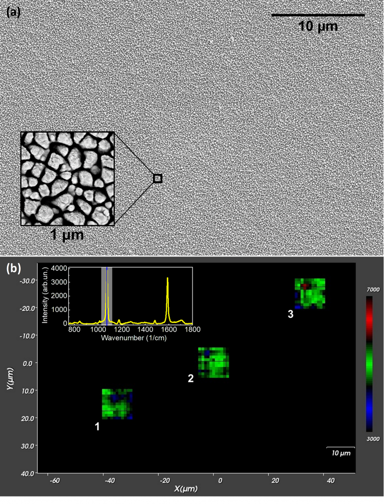
Uniformity of bSi/Au SERS substrate. (a) – Top-down SEM image of bSi/Au substrate, inset gives 1×1 μm area. (b) – Map of the background-corrected Raman intensity. 1077 cm-1 C–S stretch vibration peak, a 50× objective (NA 1.0) were used. The map resolution is 1 μm. 1,2,3 – separate 10×10 μm maps taken randomly from a 75×115 μm area. Inset gives a SERS spectrum of 4-MBA monolayer from 1×1 μm pixel.
Group currently performs theoretical modeling and experimental verification of using photonic materials of reduced dimensionality for combining medical diagnosing and treatment in vitro. This approach is often dubbed “theranostics”.
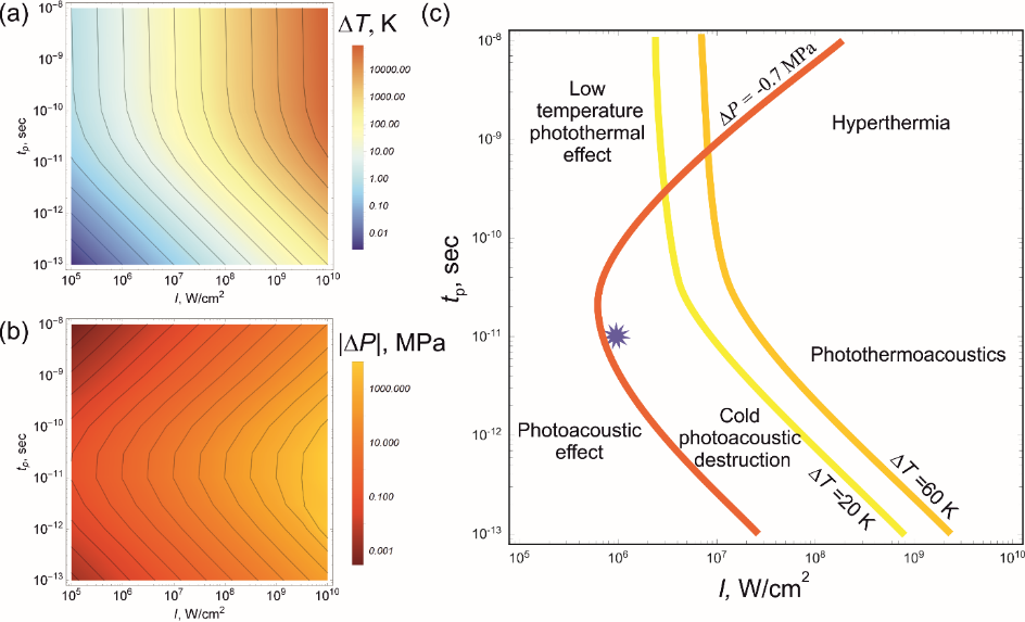
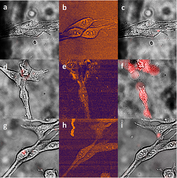
Single wall carbon nanotubes were proved to be effective theranostic agent, suitable for both detection of the cancer cells and their treatment non-invasive for surrounded healthy cells though cold photoacoustic mechanism.
Our ambition is to create and validate in vitro a simple, robust and scalable multimodal quantum theranostic agents – fluorescent nanodiamonds and single-crystalline diamond needles embedding color centers, and graphene and other 2D materials-based quantum dots – capable of monitoring the functional state of electrically active cells, i.e. neurons, and destroying cancer cells.
In research, the group actively collaborates with international partners from Center for Physical Sciences and Technology, Vilnius, Lithuania; Ulm University/ Institute for Quantum Optics, Ulm, Germany; Tor Vergata University/Department of Physics, Rome, Italy; University of Warsaw/ Quantum Optics Lab, Warsaw, Poland; Institute of Physics, National Academy of Science, Minsk, Belarus
Contact persons:
Prof. Polina Kuzhir
Prof. Yuri Svirko
Dr. Sergei Malykhin
Prof. Alexander Obraztsov
Selected publications:
- L. Golubewa, I. Timoshchenko, O. Romanov, R. Karpicz, T. Kulahava, D. Rutkauskas, M. Shuba, A. Dementjev, Y. Svirko, and P. Kuzhir, “Single-walled carbon nanotubes as a photo-thermo-acoustic cancer theranostic agent: theory and proof of the concept experiment”, Sci. Rep. 10, 22174 (2020).
- L. Golubewa, et. al., “Rapid and delayed effects of single-walled carbon nanotubes in glioma cells”, Nanotechnology 32 505103 (2021).
- L. Golubewa, et. al., “All-Optical Thermometry with NV and SiV Color Centers in Biocompatible Diamond Microneedles”, Adv. Optical Mater. 10, 2200631 (2022).
- Y. Padrez, et. al., “Quantitative and qualitative analysis of pulmonary arterial hypertension fibrosis using wide-field second harmonic generation microscopy”, Scientific Reports 12, 7330 (2022).
- N. Belko, et. al., “Hysteresis and Stochastic Fluorescence by Aggregated Ensembles of Graphene Quantum Dots”, J. Phys. Chem. C 126, 10469 (2022).
- Y. Padrez, et. al. “Nanodiamond surface as a photoluminescent pH sensor”, Nanotechnology 34 195702 (2023).
- M. Quarshie, et. al., “Nano- and micro-crystalline diamond film structuring with electron beam lithography mask”, Nanotechnology 35 155301 (2024).
Keywords:
Nanodiamonds, color center, carbon nanotube, graphene quantum dot, theranostic agent

Numerical simulation of the interaction of laser radiation with the SWCNT agglomerate embedded into the living cell. Contour maps of the a temperature increase and b negative pressure in the SWCNT agglomerate of 1 µm aggregated inside the glioma cell on the pulse duration/intensity plane. c Isotherms at ΔT = 20 K and ΔT = 60 K, respectively, and isobar at ΔP = – 0.7 MPa. The star corresponds to the experimental conditions.

Photo-induced SWCNT-mediated destruction of glioma cells by NIR pico-second pulsed irradiation. a-c Bare C6 glioma cells; d-f C6 glioma cells with accumulated micron-sized SWCNT agglomerates, g-i C6 glioma cells in the presence of the SWCNTs suspension in the extracellular medium. a, d, g bright-field images superimposed with PI fluorescence images before irradiation; b, e, h CARS images; c, f, i bright-field images superimposed with PI fluorescence images after irradiation with 10 ps laser pulses (910.5/1064 nm, 106 W/cm2) for 7 min.
Unique features of atomic-sized objects or defects allow optical quantum sensing and bioimaging. We investigate diamond nanoparticles, films and needles enriched with various types of color centers as well as quantum dots for these purposes. Sensing based on all-optical and spin approaches as well as their combination is employed. Applying nanofabrication techniques to fluorescence diamonds we create and characterize sensing matrixes and chips.

Scheme of spin-based sensing with NV centers
Publications:
10.1002/adom.202200631
10.1088/1361-6528/ad18e9
Contact persons:
Prof. Polina Kuzhir
Dr. Sergei Malykhin
Prof. Alexander Obraztsov
Other optical measurements
Sm4rtlab is totally new, revolutionary augmented reality environment combining science and teaching.
Read more about Sm4rtlab.